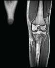
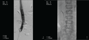
Figure 3
Bacteremia with the Triad Osteomyelitis, Deep Vein Thrombosis, and Pulmonary Septic Emboli in Pediatric Age: A Case Report
Marisol Holanda Pena*, Maria Hermoso Diez, Elsa Ots Ruiz, Ana de Berrazueta Sanchez de Vega and Jose Manuel Lanza Gomez
Published: 25 October, 2025 | Volume 9 - Issue 1 | Pages: 004-008
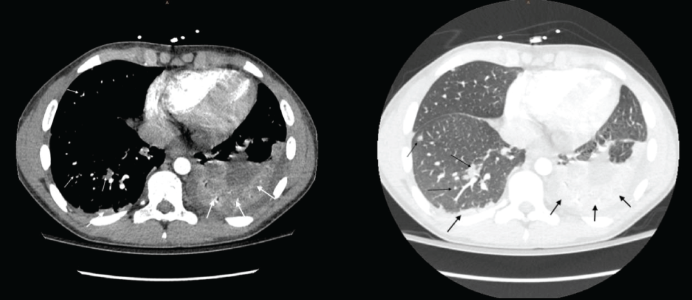
Figure 3:
CT scan showing a small bilateral pleural effusion slightly larger on the left side with passive atelectasis of both pulmonary bases (thick arrows). Multiple bilateral nodular lesions, some of them cavitated, in relation to septic embolisms with an associated pulmonary infarction (thin arrows).
Read Full Article HTML DOI: 10.29328/journal.aceo.1001022 Cite this Article Read Full Article PDF
More Images
Similar Articles
-
Bacteremia with the Triad Osteomyelitis, Deep Vein Thrombosis, and Pulmonary Septic Emboli in Pediatric Age: A Case ReportMarisol Holanda Pena*,Maria Hermoso Diez,Elsa Ots Ruiz,Ana de Berrazueta Sanchez de Vega,Jose Manuel Lanza Gomez. Bacteremia with the Triad Osteomyelitis, Deep Vein Thrombosis, and Pulmonary Septic Emboli in Pediatric Age: A Case Report. . 2025 doi: 10.29328/journal.aceo.1001022; 9: 004-008
Recently Viewed
-
Internet Addiction and its Relationship with Attachment Styles Among Tunisian Medical StudentsRim Masmoudi*, Ahmed Mhalla, Amjed Ben Haouala, Wael Majdoub, Jawaher Masmoudi, Badii Amamou, Lotfi Gaha. Internet Addiction and its Relationship with Attachment Styles Among Tunisian Medical Students. J Addict Ther Res. 2023: doi: 10.29328/journal.jatr.1001027; 7: 012-018
-
Differences between anorexia patients and participants of the Minnesota hunger experiment: Consequences for treatmentGreta Noordenbos*. Differences between anorexia patients and participants of the Minnesota hunger experiment: Consequences for treatment. J Addict Ther Res. 2021: doi: 10.29328/journal.jatr.1001013; 5: 001-002
-
Estimation of cotinine level among the tobacco users and nonusers: A cross-sectional study among the Indian populationSukhvinder Singh Oberoi*,Avneet Oberoi. Estimation of cotinine level among the tobacco users and nonusers: A cross-sectional study among the Indian population. J Addict Ther Res. 2021: doi: 10.29328/journal.jatr.1001014; 5: 003-008
-
Drug treatment and rehabilitation in China: Theoretical rationales and current situationsGloria Yuxuan Gu*. Drug treatment and rehabilitation in China: Theoretical rationales and current situations. J Addict Ther Res. 2021: doi: 10.29328/journal.jatr.1001015; 5: 009-011
-
Patterns of drugs and alcohol abuse among youthTamar Ruth Orowitz*. Patterns of drugs and alcohol abuse among youth. J Addict Ther Res. 2021: doi: 10.29328/journal.jatr.1001016; 5: 012-013
Most Viewed
-
Feasibility study of magnetic sensing for detecting single-neuron action potentialsDenis Tonini,Kai Wu,Renata Saha,Jian-Ping Wang*. Feasibility study of magnetic sensing for detecting single-neuron action potentials. Ann Biomed Sci Eng. 2022 doi: 10.29328/journal.abse.1001018; 6: 019-029
-
Evaluation of In vitro and Ex vivo Models for Studying the Effectiveness of Vaginal Drug Systems in Controlling Microbe Infections: A Systematic ReviewMohammad Hossein Karami*, Majid Abdouss*, Mandana Karami. Evaluation of In vitro and Ex vivo Models for Studying the Effectiveness of Vaginal Drug Systems in Controlling Microbe Infections: A Systematic Review. Clin J Obstet Gynecol. 2023 doi: 10.29328/journal.cjog.1001151; 6: 201-215
-
Causal Link between Human Blood Metabolites and Asthma: An Investigation Using Mendelian RandomizationYong-Qing Zhu, Xiao-Yan Meng, Jing-Hua Yang*. Causal Link between Human Blood Metabolites and Asthma: An Investigation Using Mendelian Randomization. Arch Asthma Allergy Immunol. 2023 doi: 10.29328/journal.aaai.1001032; 7: 012-022
-
Impact of Latex Sensitization on Asthma and Rhinitis Progression: A Study at Abidjan-Cocody University Hospital - Côte d’Ivoire (Progression of Asthma and Rhinitis related to Latex Sensitization)Dasse Sery Romuald*, KL Siransy, N Koffi, RO Yeboah, EK Nguessan, HA Adou, VP Goran-Kouacou, AU Assi, JY Seri, S Moussa, D Oura, CL Memel, H Koya, E Atoukoula. Impact of Latex Sensitization on Asthma and Rhinitis Progression: A Study at Abidjan-Cocody University Hospital - Côte d’Ivoire (Progression of Asthma and Rhinitis related to Latex Sensitization). Arch Asthma Allergy Immunol. 2024 doi: 10.29328/journal.aaai.1001035; 8: 007-012
-
An algorithm to safely manage oral food challenge in an office-based setting for children with multiple food allergiesNathalie Cottel,Aïcha Dieme,Véronique Orcel,Yannick Chantran,Mélisande Bourgoin-Heck,Jocelyne Just. An algorithm to safely manage oral food challenge in an office-based setting for children with multiple food allergies. Arch Asthma Allergy Immunol. 2021 doi: 10.29328/journal.aaai.1001027; 5: 030-037

If you are already a member of our network and need to keep track of any developments regarding a question you have already submitted, click "take me to my Query."


