Figure 1
Bacteremia with the Triad Osteomyelitis, Deep Vein Thrombosis, and Pulmonary Septic Emboli in Pediatric Age: A Case Report
Marisol Holanda Pena*, Maria Hermoso Diez, Elsa Ots Ruiz, Ana de Berrazueta Sanchez de Vega and Jose Manuel Lanza Gomez
Published: 25 October, 2025 | Volume 9 - Issue 1 | Pages: 004-008
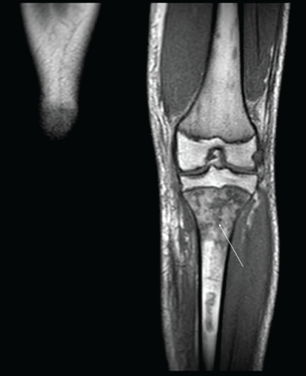
Figure 1:
MRI of the knee joint. In the proximal metaphysis and diaphysis of the left tibia, there is a marked alteration in signal intensity, which appears hyperintense at T2 and hypointense at T1, with multiple well-defined focal lesions of serpiginous and irregular morphology suggestive of multiple bone infarctions in the context of osteomyelitis.
Read Full Article HTML DOI: 10.29328/journal.aceo.1001022 Cite this Article Read Full Article PDF
More Images
Similar Articles
-
Bacteremia with the Triad Osteomyelitis, Deep Vein Thrombosis, and Pulmonary Septic Emboli in Pediatric Age: A Case ReportMarisol Holanda Pena*,Maria Hermoso Diez,Elsa Ots Ruiz,Ana de Berrazueta Sanchez de Vega,Jose Manuel Lanza Gomez. Bacteremia with the Triad Osteomyelitis, Deep Vein Thrombosis, and Pulmonary Septic Emboli in Pediatric Age: A Case Report. . 2025 doi: 10.29328/journal.aceo.1001022; 9: 004-008
Recently Viewed
-
Internet Addiction and its Relationship with Attachment Styles Among Tunisian Medical StudentsRim Masmoudi*, Ahmed Mhalla, Amjed Ben Haouala, Wael Majdoub, Jawaher Masmoudi, Badii Amamou, Lotfi Gaha. Internet Addiction and its Relationship with Attachment Styles Among Tunisian Medical Students. J Addict Ther Res. 2023: doi: 10.29328/journal.jatr.1001027; 7: 012-018
-
Differences between anorexia patients and participants of the Minnesota hunger experiment: Consequences for treatmentGreta Noordenbos*. Differences between anorexia patients and participants of the Minnesota hunger experiment: Consequences for treatment. J Addict Ther Res. 2021: doi: 10.29328/journal.jatr.1001013; 5: 001-002
-
Estimation of cotinine level among the tobacco users and nonusers: A cross-sectional study among the Indian populationSukhvinder Singh Oberoi*,Avneet Oberoi. Estimation of cotinine level among the tobacco users and nonusers: A cross-sectional study among the Indian population. J Addict Ther Res. 2021: doi: 10.29328/journal.jatr.1001014; 5: 003-008
-
Drug treatment and rehabilitation in China: Theoretical rationales and current situationsGloria Yuxuan Gu*. Drug treatment and rehabilitation in China: Theoretical rationales and current situations. J Addict Ther Res. 2021: doi: 10.29328/journal.jatr.1001015; 5: 009-011
-
Patterns of drugs and alcohol abuse among youthTamar Ruth Orowitz*. Patterns of drugs and alcohol abuse among youth. J Addict Ther Res. 2021: doi: 10.29328/journal.jatr.1001016; 5: 012-013
Most Viewed
-
Feasibility study of magnetic sensing for detecting single-neuron action potentialsDenis Tonini,Kai Wu,Renata Saha,Jian-Ping Wang*. Feasibility study of magnetic sensing for detecting single-neuron action potentials. Ann Biomed Sci Eng. 2022 doi: 10.29328/journal.abse.1001018; 6: 019-029
-
Evaluation of In vitro and Ex vivo Models for Studying the Effectiveness of Vaginal Drug Systems in Controlling Microbe Infections: A Systematic ReviewMohammad Hossein Karami*, Majid Abdouss*, Mandana Karami. Evaluation of In vitro and Ex vivo Models for Studying the Effectiveness of Vaginal Drug Systems in Controlling Microbe Infections: A Systematic Review. Clin J Obstet Gynecol. 2023 doi: 10.29328/journal.cjog.1001151; 6: 201-215
-
Causal Link between Human Blood Metabolites and Asthma: An Investigation Using Mendelian RandomizationYong-Qing Zhu, Xiao-Yan Meng, Jing-Hua Yang*. Causal Link between Human Blood Metabolites and Asthma: An Investigation Using Mendelian Randomization. Arch Asthma Allergy Immunol. 2023 doi: 10.29328/journal.aaai.1001032; 7: 012-022
-
Impact of Latex Sensitization on Asthma and Rhinitis Progression: A Study at Abidjan-Cocody University Hospital - Côte d’Ivoire (Progression of Asthma and Rhinitis related to Latex Sensitization)Dasse Sery Romuald*, KL Siransy, N Koffi, RO Yeboah, EK Nguessan, HA Adou, VP Goran-Kouacou, AU Assi, JY Seri, S Moussa, D Oura, CL Memel, H Koya, E Atoukoula. Impact of Latex Sensitization on Asthma and Rhinitis Progression: A Study at Abidjan-Cocody University Hospital - Côte d’Ivoire (Progression of Asthma and Rhinitis related to Latex Sensitization). Arch Asthma Allergy Immunol. 2024 doi: 10.29328/journal.aaai.1001035; 8: 007-012
-
An algorithm to safely manage oral food challenge in an office-based setting for children with multiple food allergiesNathalie Cottel,Aïcha Dieme,Véronique Orcel,Yannick Chantran,Mélisande Bourgoin-Heck,Jocelyne Just. An algorithm to safely manage oral food challenge in an office-based setting for children with multiple food allergies. Arch Asthma Allergy Immunol. 2021 doi: 10.29328/journal.aaai.1001027; 5: 030-037

If you are already a member of our network and need to keep track of any developments regarding a question you have already submitted, click "take me to my Query."


