
More Information
Submitted: October 06, 2023 | Approved: November 01, 2023 | Published: November 02, 2023
How to cite this article: Castagno C, Campano D, Fernandez I, Weiss WM. Masquelet Technique for Reconstruction of a Fourth Metatarsal Defect Following a Low-velocity Gunshot Wound: A Case Report. Arch Clin Exp Orthop. 2023; 7: 022-026.
DOI: 10.29328/journal.aceo.1001017
Copyright Licence: © 2023 Castagno C, et al. This is an open access article distributed under the Creative Commons Attribution License, which permits unrestricted use, distribution, and reproduction in any medium, provided the original work is properly cited.
Keywords: Masquelet; Induced-membrane; Pseudomembrane; Pseudosynovial membrane; Reconstruction; Fourth metatarsal; Gunshot wound; PMMA space

Masquelet Technique for Reconstruction of a Fourth Metatarsal Defect Following a Low-velocity Gunshot Wound: A Case Report
Castagno C1*, Campano D2, Fernandez I2 and Weiss WM3
1Texas Tech University Health Science Center, Paul Foster School of Medicine, El Paso, TX, USA
2Orthopedic Surgery & Sports Medicine, William Beaumont Army Medical Center, Fort Bliss, TX, USA
3Orthopedic Surgery & Sports Medicine, University of Texas Medical Branch, Galveston, TX, USA
*Address for Correspondence: Castagno C, Texas Tech University Health Science Center, Paul Foster School of Medicine, El Paso, 4815 Alameda Ave. El Paso, TX 79905, USA, Email: [email protected]
The Masquelet technique has become increasingly popular in reconstruction scenarios such as osteomyelitis, cancer, and nonunions. The procedure is a two-staged approach: first, a polymethylmethacrylate (PMMA) cement spacer is inserted to fill a bone void. The spacer induces a membrane to form around it, and 6-9 weeks later, the second stage involves carefully extracting the spacer and filling the membrane with bone graft. Most of the current literature has published either femoral or tibial involvement for Masquelet studies, with limited published data on foot reconstruction. Here, the authors used the procedure for the fourth metatarsal. Despite complications, this case proves a more excellent utility and warrants further investigation into Masquelet techniques in the foot.
Gunshot injuries are common in combat situations but are now a growing problem in the civilian population of the United States. Between 2010 and 2012, greater than 32,000 people per year were killed by firearms, with the majority being suicides [1]. Of those fatalities, an average of 582 a year were accidental [1]. Over the same time frame, there were 11,529 unintentional firearm injuries yearly [1]. Of this group, 55% were younger than 34. The economic impact of gunshot wounds is a median cost of $56,000 per hospitalization, and this does not account for time away from work and recovery for these young patients [2,3]. Prognosis and return to work or activity following gunshot injuries are highly dependent on the nature of the damage and can range from complete recovery to disability. Gunshot injuries to the extremities can result in significant soft tissue and bone defects that require advanced reconstruction techniques or amputation.
The Masquelet technique, introduced in 1986, has become increasingly popular for reconstruction scenarios with bone defects, such as those encountered in trauma, osteomyelitis, and cancer. The technique allows restoration of up to 30% - 50% of bone length using an induced membrane and bone graft [4]. It is a two-step procedure, the first of which places a cement spacer in the bone defect, which induces the formation of a biological membrane surrounding it. The induced membrane forms a contained space for subsequent bone grafting and an environment that promotes healing. Outcomes have been successful, with clinical and radiographic union rates of up to 90% in one year [5]. Classically described in the femur and tibia, three but has also been described in the clavicle, fibula, ulna, and humerus [6-9]. It has demonstrated effectiveness in adult and pediatric populations, managing congenital pseudoarthrosis and tumor resections [9-12]. The Masquelet technique has been described in the first metatarsal by Makridis, et al. in Foot Ankle Surgery published in 2014. Still, there needs to be literature describing the use of this technique in the lesser metatarsals.
This case explores the application of the Masquelet technique to the foot to treat bone loss from the fourth metatarsal bone following an accidental close-range low-velocity gunshot wound. The patient consented to the publication of all information disclosed in this report, non-pertinent patient information was not disclosed, and all ethical considerations were reviewed by the authors.
A healthy 29-year-old female with no pertinent PMH sustained an accidental self-inflicted close-range, low-velocity gunshot wound to the left foot. Physical exam was significant for a 10 x 25 mm dorsal entry wound and 20 x 30 mm plantar exit wound, with retained metallic fragments. Radiographs demonstrated a transverse displaced third metatarsal diaphysis fracture, segmental bone loss of about 15 mm between the head and diaphysis of the fourth metatarsal, and a minimally displaced fifth metatarsal base fracture. Injuries were isolated to the left foot. The fourth toe was pink and perfused but had an altered sensation. The remainder of the neurovascular examination of the foot was ordinary.
Initial management of the Gustilo II wound included radiographic examination, irrigation, and debridement in the emergency room (Figure 1). The patient’s tetanus status was confirmed as up to date, and antibiotics (clindamycin due to a penicillin allergy) were started. The foot was dressed and splinted in preparation for formal treatment in the operating room the next day. Antibiotic prophylaxis was again given before surgery, and the patient underwent standard irrigation and debridement in the operating room the day following admission. This included the removal of metallic debris, de-vascularized bone fragments, and devitalized tissue in addition to irrigation with 6L of normal saline. With the assistance of fluoroscopy, a 0.062 Kirschner wire was placed retrograde across the 3rd metatarsal fracture after reduction. Two transverse 0.062 Kirschner wires were identified from the 5th to the 3rd metatarsal to stabilize the 5th and maintain the length of the 4th. A 10 x 15 mm molded antibiotic-eluting cement spacer (Stryker) was then placed in the defect of the 4th metatarsal as stage 1 of the Masquelet technique. The plantar and dorsal wounds were then loosely approximated with nylon sutures (Figure 2). The patient was discharged on the day of surgery, non-weight bearing in a fracture boot, to await stage 2 of the Masquelet technique.
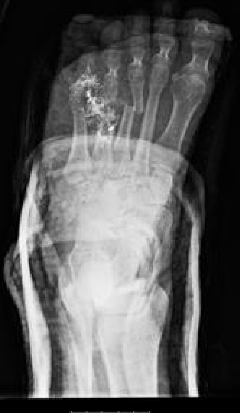
Figure 1: XR of the left foot after gross irrigation and debridement, splint placement; note the metallic debris over the 4th metatarsal.
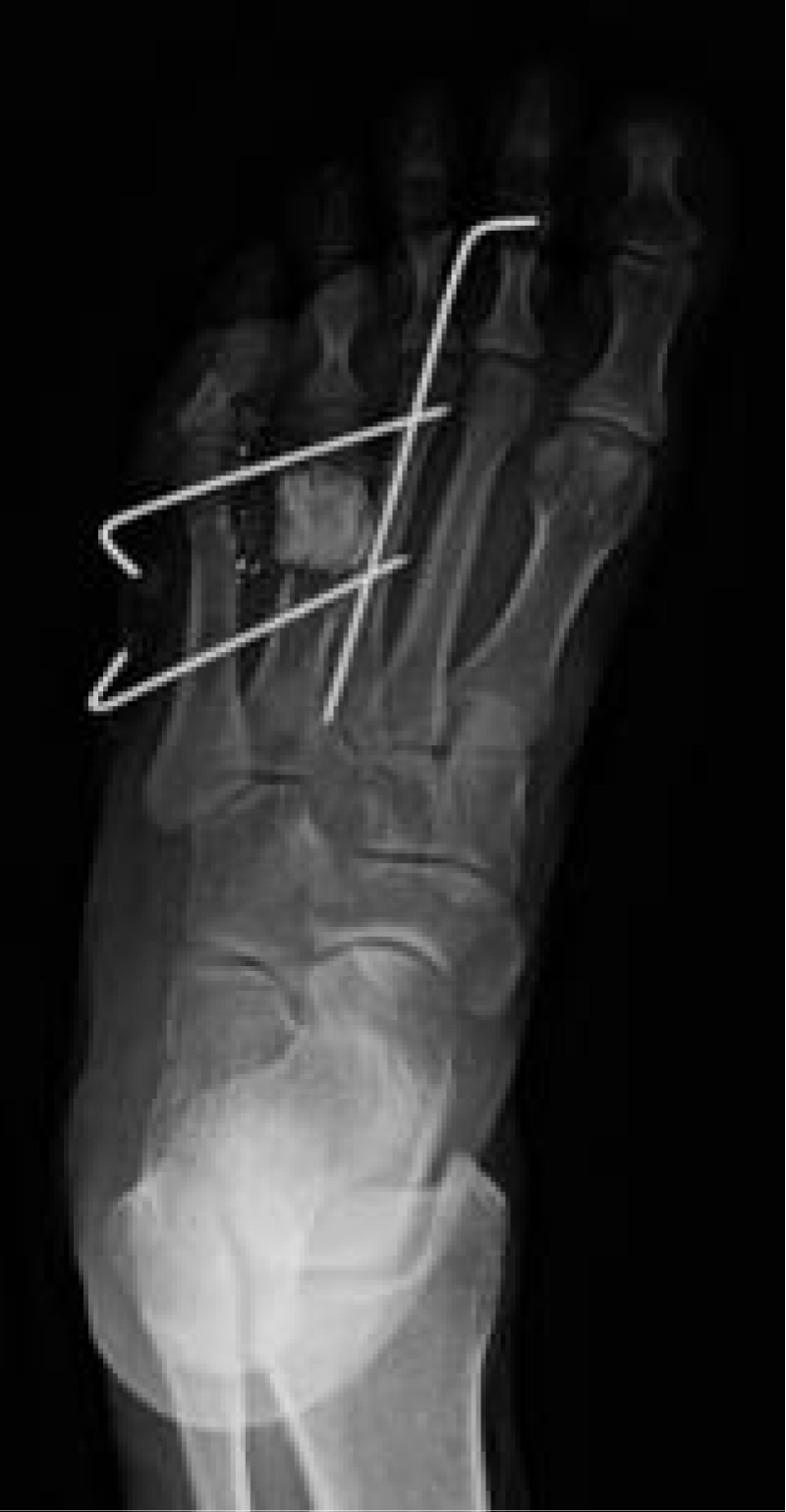
Figure 2: Transverse pins are placed to hold the 4th metatarsal out to length. The PMMA spacer is visibly occupying the bone loss.
The patient was seen at two weeks for wound check and suture removal, with no evidence of infection or wound breakdown. At six weeks, radiographs demonstrated the reduction was maintained, with the hardware and cement spacer in a good position with no evidence of failure. Pins were removed, and the patient was instructed to continue non-weight bearing in the fracture boot. At about eight weeks postoperatively, the patient presented with wound dehiscence dorsally and exposed cement spacer. The cement spacer was removed in the clinic, and the patient was placed on oral antibiotics and dressing changes every two days until the wound was closed (Figure 3).
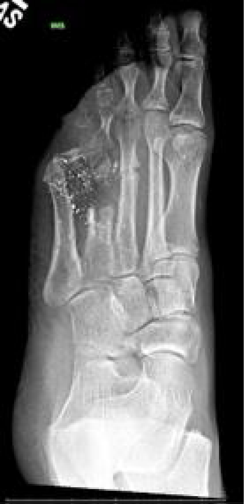
Figure 3: showing the foot after complication and spacer removal.
At 15 weeks postoperatively, the patient’s dorsal wound had closed, and stage 2 of the Masquelet procedure was performed. At the time of surgery, a thick pseudomembrane was encountered where the cement spacer had been placed between the 4th metatarsal head and diaphysis, deep to the healed skin defect. The pseudomembrane was incised in line with the skin incision over the defect. A fibular allograft (Depuy) was fashioned to fit the fault. Of note, some shortening had occurred while there was no longer a cement spacer to maintain the length of the 4th ray due to soft tissue contracture, but the construct provided a 4th ray that was only slightly longer than the 5th. A 7-hole low profile plate (Arthrex) was placed, and excellent purchase was obtained in the head and diaphysis to secure the allograft between them. The pseudomembrane was then closed over the construct with an absorbable suture, followed by the skin (Figure 4). The patient was placed in a non-weight bearing short leg cast for six weeks.
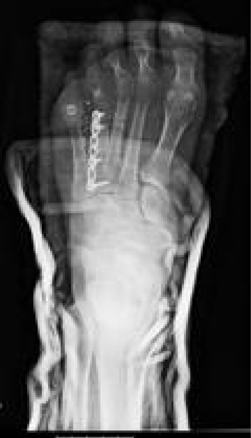
Figure 4: 7-hole Arthrex plate and fibular graft.
At six weeks postoperatively from stage 2 of the Masquelet technique, the patient was transitioned into a fracture boot and began weight bearing as tolerated. At 16 weeks from stage 2, the patient was transitioned to a regular shoe and began conditioning for return to work. At seven months from stage 1 of the Masquelet procedure, the patient had returned to complete work with no difficulties but was not yet running. At nine months from stage 1, the patient performed all work duties and could run without problem (Figure 5).
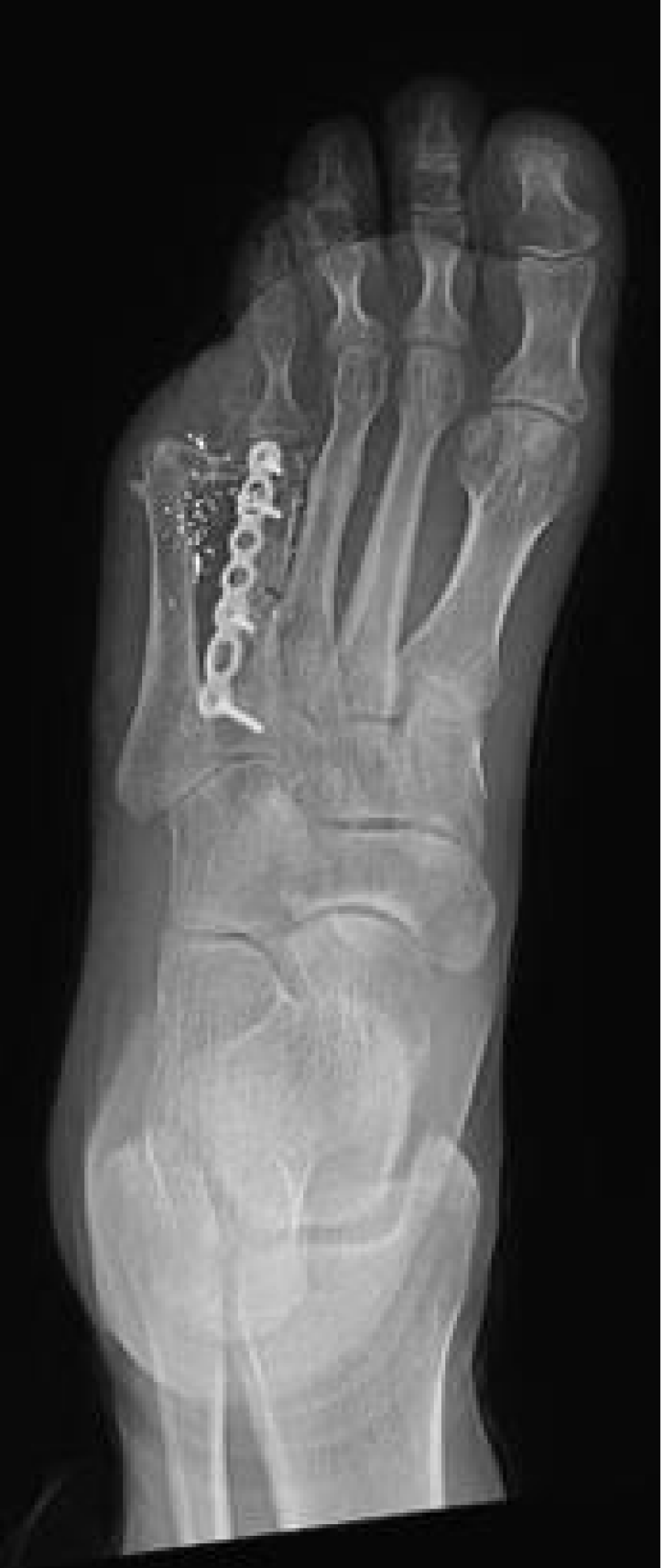
Figure 5: XR reveals intact hardware with no evidence of failure.
This case report highlights the ability of the Masquelet technique to overcome a bone defect and maintain metatarsal length despite the need to remove the cement spacer before the procedure’s second stage. In the second stage, the thick pseudomembrane was still present and surrounded the void left by the bone defect. As noted in the literature, this membrane is highly vascularized, providing a responsive bone growth environment [13-15]. Successful union was achieved, and the patient could return to active whole duty without pain or problems. This is the only successful use of the Masquelet technique with spacer removal before the second stage and the only point in the lesser metatarsals.
Results of the Masquelet technique in the femur and tibia have been well characterized, as Masquelet, et al. published a 35-patient case series with an average time to union of 8.5 months. An average time to unrestricted weight-bearing has been reported at 7.9 months from stage 1 and 4.5 months from stage 2, amongst tibial and femoral cases [14-16]. In keeping with this, union and full weight bearing were achieved in our case of the metatarsal Masquelet technique in 7 months from the index procedure.
Cases of using the Masquelet technique in the foot have been reported, but none in the lesser metatarsals. Liu, et al. report using the Masquelet technique to treat first metatarsophalangeal joint gout with bone defect; results were favorable—total combined excellent and reasonable rate was 63.6% [17,18]. However, the treatment of gout in this manner is controversial, and it is unclear if the Masquelet technique was successful as the patient underwent more extensive surgery, including extensive arthrodesis [15]. The authors stabilized the first metatarsal fracture with a mini-external fixator and bridged the bone defect with two locking plates. At ten months postoperatively, residual pain and instability led to arthrodesis of the first MTP joint combined with hindfoot and tibiotalar joint. Largey, et al. documented a case with medial cuneiform bone loss managed with a Masquelet and a saphenous cross-leg flap [17-19].
Complications of the induced-membrane technique previously reported include nonunion, wound healing issues, amputation, and fistula [18-20]. Emphasis on a thorough debridement ensures a healthy pseudomembranous environment and minimizes infection risk [19]. Relative contraindications to this technique affect bone’s healing ability, including tissue necrosis, radiation, chronic systemic steroid use, smoking, and malnutrition [19-24]. While the patient had a dorsal wound that re-opened following initial close closure, the wound did close by secondary intention following removal of the spacer before the second stage. Despite removing the cement spacer, the pseudomembrane remained intact until the second stage, which current literature suggests has never been previously reported.
Alternatives to the treatment described included amputation of the 4th toe. However, this is likely somewhat aggressive without evidence of the patient’s inability to tolerate the limited function of the 4th toe in this state. Another option is a free fibular autograft, described in metatarsal defects, though usually for those larger than 6 cm [25]. The shape of the fibula, ease of harvest, and donor site vessel diameter are advantages when using this technique. However, donor site pain, duration of surgery, and the need for a specialist for vessel anastomosis make this a less attractive option, particularly for a relatively small bone defect. The simplest option is to leave the bony defect as it was and hope for a fibrous union to bridge the metatarsal gap. But this would likely have resulted in the shortening of the 4th ray and an unstable union that would effectively eliminate the 4th toe in weight-bearing and result in a chronic metatarsalgia of the 3rd and 5th metatarsal heads due to overloading.
Limitations of the current investigation include the inherent limitations of an isolated case study. While the patient has achieved clinical union, radiographic union is difficult to define and demonstrate. Shortening the 4th metatarsal is not ideal, and while the patient is doing well, the possibility remains of developing metatarsalgia at the heads of the 3rd or 5th.
The Masquelet technique has been demonstrated to be effective in the foot, and this case report represents the first published case of its use in the lesser metatarsals and demonstrates the technique’s effectiveness despite removing the cement spacer before the second stage. It remains an effective method for managing bone loss in traumatic situations and may allow for early spacer removal if required.
- Fowler KA, Dahlberg LL, Haileyesus T, Annest JL. Firearm injuries in the United States. Prev Med. 2015 Oct;79:5-14. doi: 10.1016/j.ypmed.2015.06.002. Epub 2015 Jun 24. PMID: 26116133; PMCID: PMC4700838.
- Cook A, Osler T, Hosmer D, Glance L, Rogers F, Gross B, Garcia-Filion P, Malhotra A. Gunshot wounds resulting in hospitalization in the United States: 2004-2013. Injury. 2017 Mar;48(3):621-627. doi: 10.1016/j.injury.2017.01.044. Epub 2017 Jan 30. PMID: 28173921..
- Bartlett CS, Helfet DL, Hausman MR, Strauss E. Ballistics and gunshot wounds: effects on musculoskeletal tissues. J Am Acad Orthop Surg. 2000 Jan-Feb;8(1):21-36. doi: 10.5435/00124635-200001000-00003. PMID: 10666650.
- Gouron R. Surgical technique and indications of the induced membrane procedure in children. Orthop Traumatol Surg Res. 2016 Feb;102(1 Suppl):S133-9. doi: 10.1016/j.otsr.2015.06.027. Epub 2016 Jan 7. PMID: 26774902.
- Stafford PR, Norris BL. Reamer-irrigator-aspirator bone graft and bi Masquelet technique for segmental bone defect nonunions: a review of 25 cases. Injury. 2010 Nov;41 Suppl 2:S72-7. doi: 10.1016/S0020-1383(10)70014-0. PMID: 21144933.
- Morris R, Hossain M, Evans A, Pallister I. Induced membrane technique for treating tibial defects gives mixed results. Bone Joint J. 2017 May;99-B(5):680-685. doi: 10.1302/0301-620X.99B5.BJJ-2016-0694.R2. PMID: 28455479.
- Abdellaoui H, Atarraf K, Chater L, Afifi MA. Congenital pseudarthrosis of the clavicle treated by Masquelet technique. BMJ Case Rep. 2017 Nov 8;2017:bcr2017221557. doi: 10.1136/bcr-2017-221557. PMID: 29122899; PMCID: PMC5695429.
- Calori GM, Mazza EL, Colombo A, Mazzola S, Colombo M. Treatment of an atrophic clavicle non-union with the chamber induction technique: a case report. Injury. 2017 Oct;48 Suppl 3:S71-S75. doi: 10.1016/S0020-1383(17)30662-9. PMID: 29025615.
- Morelli I, Drago L, George DA, Romanò D, Romanò CL. Managing large bone defects in children: a systematic review of the 'induced membrane technique'. J Pediatr Orthop B. 2018 Sep;27(5):443-455. doi: 10.1097/BPB.0000000000000456. PMID: 28368930.
- Konda SR, Gage M, Fisher N, Egol KA. Segmental Bone Defect Treated With the Induced Membrane Technique. J Orthop Trauma. 2017 Aug;31 Suppl 3:S21-S22. doi: 10.1097/BOT.0000000000000899. PMID: 28697078.
- Tong K, Zhong Z, Peng Y, Lin C, Cao S, Yang Y, Wang G. Masquelet technique versus Ilizarov bone transport for reconstruction of lower extremity bone defects following posttraumatic osteomyelitis. Injury. 2017 Jul;48(7):1616-1622. doi: 10.1016/j.injury.2017.03.042. Epub 2017 Apr 4. PMID: 28408083.
- Masquelet AC. Induced Membrane Technique: Pearls and Pitfalls. J Orthop Trauma. 2017 Oct;31 Suppl 5:S36-S38. doi: 10.1097/BOT.0000000000000979. PMID: 28938390.
- Makridis KG, Theocharakis S, Fragkakis EM, Giannoudis PV. Reconstruction of an extensive soft tissue and bone defect of the first metatarsal with the use of Masquelet technique: a case report. Foot Ankle Surg. 2014 Jun;20(2):e19-22. doi: 10.1016/j.fas.2013.11.006. Epub 2013 Dec 16. PMID: 24796840.
- Zoller SD, Cao LA, Smith RA, Sheppard W, Lord EL, Hamad CD, Ghodasra JH, Lee C, Jeffcoat D. Staged reconstruction of diaphyseal fractures with segmental defects: Surgical and patient-reported outcomes. Injury. 2017 Oct;48(10):2248-2252. doi: 10.1016/j.injury.2017.06.018. Epub 2017 Jun 23. PMID: 28712488.
- Liu F, Huang RK, Xie M, Pan H, Zhao JJ, Lei B. Use of Masquelet's technique for treating the first metatarsophalangeal joint in cases of gout combined with a massive bone defect. Foot Ankle Surg. 2018 Apr;24(2):159-163. doi: 10.1016/j.fas.2017.01.009. Epub 2017 Feb 17. PMID: 29409218.
- Pelissier P, Bollecker V, Martin D, Baudet J. Reconstruction du pied par la technique "bi-Masquelet" [Foot reconstruction with the "bi-Masquelet" procedure]. Ann Chir Plast Esthet. 2002 Aug;47(4):304-7. French. doi: 10.1016/s0294-1260(02)00123-1. PMID: 12420622.
- Largey A, Faline A, Hebrard W, Hamoui M, Canovas F. Management of massive traumatic compound defects of the foot. Orthop Traumatol Surg Res. 2009 Jun;95(4):301-4. doi: 10.1016/j.otsr.2009.02.005. Epub 2009 May 12. PMID: 19442599.
- Moghaddam A, Zietzschmann S, Bruckner T, Schmidmaier G. Treatment of atrophic tibia non-unions according to 'diamond concept': Results of one- and two-step treatment. Injury. 2015 Oct;46 Suppl 4:S39-50. doi: 10.1016/S0020-1383(15)30017-6. PMID: 26542865.
- Giannoudis PV, Faour O, Goff T, Kanakaris N, Dimitriou R. Masquelet technique for the treatment of bone defects: tips-tricks and future directions. Injury. 2011 Jun;42(6):591-8. doi: 10.1016/j.injury.2011.03.036. Epub 2011 May 4. PMID: 21543068.
- Micev AJ, Kalainov DM, Soneru AP. Masquelet technique for treatment of segmental bone loss in the upper extremity. J Hand Surg Am. 2015 Mar;40(3):593-8. doi: 10.1016/j.jhsa.2014.12.007. Epub 2015 Jan 31. PMID: 25648786.
- Apard T, Bigorre N, Cronier P, Duteille F, Bizot P, Massin P. Two-stage reconstruction of post-traumatic segmental tibia bone loss with nailing. Orthop Traumatol Surg Res. 2010 Sep;96(5):549-53. doi: 10.1016/j.otsr.2010.02.010. Epub 2010 Jun 4. PMID: 20605548.
- Flamans B, Pauchot J, Petite H, Blanchet N, Rochet S, Garbuio P, Tropet Y, Obert L. Pertes de substance osseuse à la main et au poignet traitées en urgence par technique de la membrane induite (technique de Masquelet) [Use of the induced membrane technique for the treatment of bone defects in the hand or wrist, in emergency]. Chir Main. 2010 Oct;29(5):307-14. French. doi: 10.1016/j.main.2010.06.008. Epub 2010 Jul 24. PMID: 20728395.
- Jiang N, Qin CH, Ma YF, Wang L, Yu B. Possibility of one-stage surgery to reconstruct bone defects using the modified Masquelet technique with degradable calcium sulfate as a cement spacer: A case report and hypothesis. Biomed Rep. 2016 Mar;4(3):374-378. doi: 10.3892/br.2016.584. Epub 2016 Jan 27. PMID: 26998279; PMCID: PMC4774369.
- Gaio N, Martino A, Toth Z, Watson JT, Nicolaou D, McBride-Gagyi S. Masquelet technique: The effect of altering implant material and topography on membrane matrix composition, mechanical and barrier properties in a rat defect model. J Biomech. 2018 Apr 27;72:53-62. doi: 10.1016/j.jbiomech.2018.02.026. Epub 2018 Feb 27. PMID: 29510858; PMCID: PMC5895482.
- Salgado CJ, Lin CH, Fuller DA, Duncan AN, Camison L, Mardini S. Foot salvage after loss of the first and second metatarsal rays with a free fibular osteocutaneous flap. J Am Podiatr Med Assoc. 2011 Nov-Dec;101(6):531-6. doi: 10.7547/1010531. PMID: 22106202.